The use of analytical balance calibration in pharmaceutical laboratories is crucial for the accurate measurement of both active substances and excipients. Its extraordinary accuracy eliminates the possibility of formulation errors and makes regulatory compliance easier. analytical balance calibration are employed by laboratory staff for daily quality control, validation of batches, and research activities. Adding analytical balance calibration to the laboratory operations not only the consistency but also the reproducibility and the accuracy of the results for clinical trials and research applications are assured.

Hospital research centers utilize analytical balance calibration in the process of making experimental medical materials and diagnostic compounds. Accurate mass measurement gives the researchers the power to supervise the formulation ratios during the first evaluation stage. This technology promotes organized experimentation, reproducibility and performance assessment among different research samples. analytical balance calibration is making the trustworthiness of experimental workflows in medical innovation environments by supplying the consistent mass data.
The era of analytical balance calibration in hospitals will go beyond the traditional settings and embrace multidisciplinary research environments. As the partnership of clinical, pharmaceutical, and biomedical teams becomes more robust, the analytical balances will cater to different experimental needs. By taking on various analytical actions, analytical balance calibration will still be a fundamental tool in contemporary hospital laboratory ecosystems.
One of the main tasks in the maintenance of analytical balance calibration in the hospital laboratory is monitoring the environmental exposure. The presence of excess humidity, direct sunlight, and temperature changes should be completely ruled out. Draft shields should always be kept in a clean and working condition to cause the least possible disturbance in air during the process of weighing. These preventive activities not only help to achieve stable measurements but also aid to lessen the variability in analytical data coming from different medical testing environments.
Medical research laboratories rely on analytical balance calibration to determine the weight of samples for their experimental procedures. No matter if weighing chemicals, biomolecules, or powders, accuracy is vital for repeatability. Scientific workers apply analytical balance calibration so that slight changes in sample weight do not affect the validity of the results. The application of this tool helps to enhance the reputation of the laboratory, the quality of the experiments, and the uniformity of the research conducted in hospitals or drug companies.
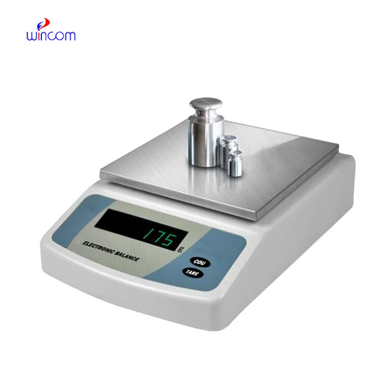
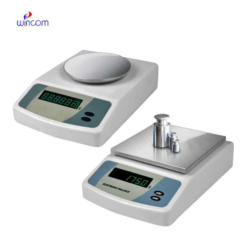
Q: What is the main purpose of an Analytical Balance? A: Its purpose is mainly to measure very tiny sample masses with the utmost precision in laboratories and hospitals. Q: What is the typical weighing range of an Analytical Balance? A: The weighing range for the majority of analytical balances is from 0 up to some grams with a resolution of micrograms or milligrams. Q: What environmental controls are necessary for an Analytical Balance's operation? A: Airflow, vibration, and temperature changes should not only be avoided but also prevented in the room where the scale is situated. Q: Is an Analytical Balance permitted in a hospital laboratory? A: Yes, it has indeed found widespread usage for the preparation of reagents, calibra¬tion, and drug development applications. Q: What should be the frequency of calibration for an Analytical Balance? A: The calibration interval is subject to the degree of use and the particular laboratory requirements.
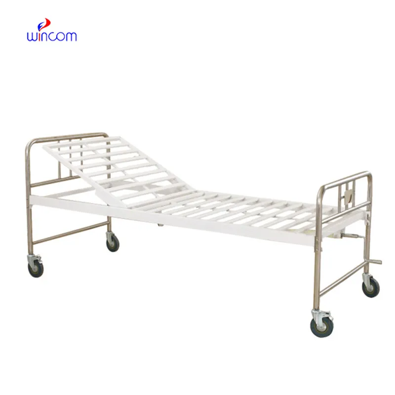
The hospital bed is well-designed and very practical. Patients find it comfortable, and nurses appreciate how simple it is to operate.
The microscope delivers incredibly sharp images and precise focusing. It’s perfect for both professional lab work and educational use.
To protect the privacy of our buyers, only public service email domains like Gmail, Yahoo, and MSN will be displayed. Additionally, only a limited portion of the inquiry content will be shown.
Could you please provide more information about your microscope range? I’d like to know the magnif...
We are planning to upgrade our imaging department and would like more information on your mri machin...
E-mail: [email protected]
Tel: +86-731-84176622
+86-731-84136655
Address: Rm.1507,Xinsancheng Plaza. No.58, Renmin Road(E),Changsha,Hunan,China

 af
af
 es
es
 ar
ar
 tr
tr
 sw
sw
 pt
pt
 th
th
 ur
ur
 bn
bn
 ne
ne
 vi
vi
 km
km
 lo
lo
 de
de
 ru
ru
 fi
fi
 nl
nl
 fa
fa
 fr
fr
 ko
ko