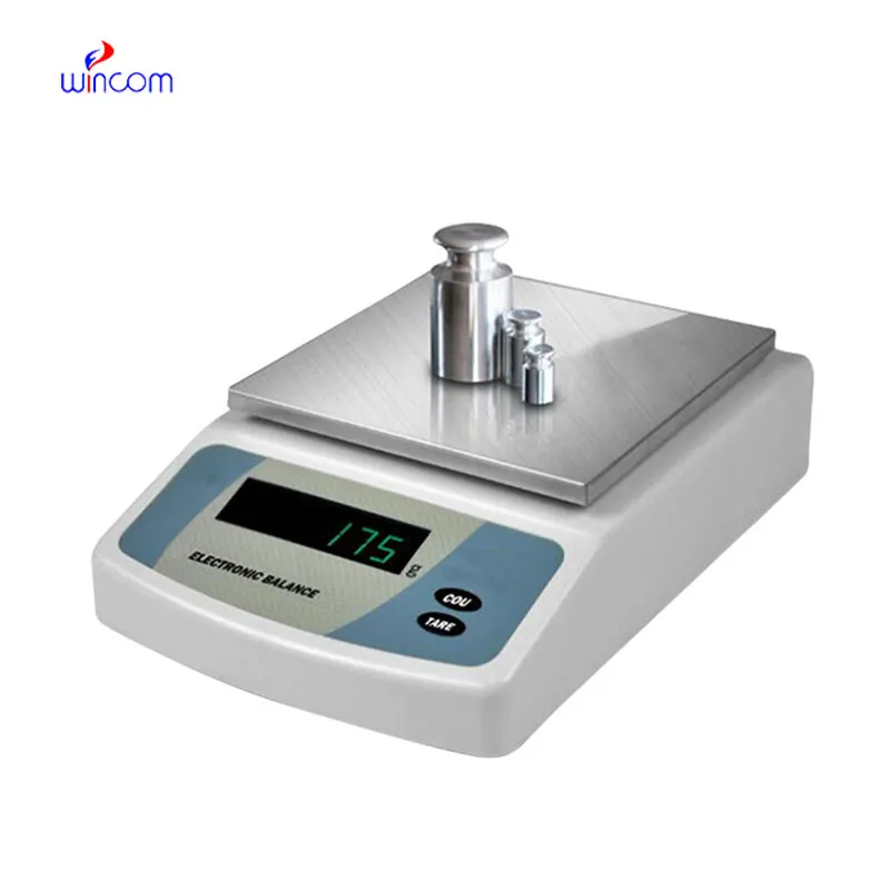
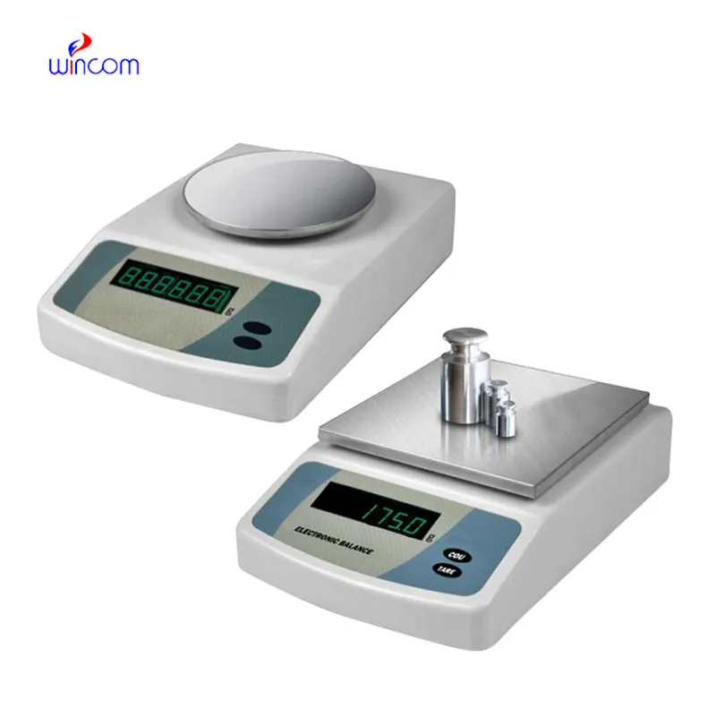
electronic balance used for ensures accurate weighing of clinical sample preparation, research studies, and hospital and laboratory medication formulation. It has an application envisaged to measure even the tiniest amounts with excellent sensitivity and repeatability. electronic balance used for is the instrument laboratory technicians trust for maintaining the highest level of accuracy in analysis, validation of experimental practices, and patients' care support. The use of this instrument in the lab operations not only guarantees results that are reliable but also creates a consistent workflow and quality control which is improved in both the diagnostic and research environments.

Hospitals' analytical laboratories use electronic balance used for during the production of internal standards for instrument testing methods. Very accurate mass input is a must to maintain uniformity over different analytical runs. This use case allows for comparison of still data, traceability, and monitoring of analytical performance over a long period. By allowing exact preparation of reference materials, electronic balance used for boosts measurement confidence in all phases of hospital laboratory work.
electronic balance used for will envision a new era where the user-centered interfaces of hospital workers take greater priority. Such friendly visualizations, accompanied by the directing of workflows and notifications, will prove to be helpful during the working of professionals with machines. This change will cut down the duration of training and will also bring in better quality of work in clinical laboratories. electronic balance used for will not give up on the accuracy aspect while still allowing the ease of use in the hospital environments that are changing fast.
The manner in which samples are handled is of utmost importance in the preservation of electronic balance used for. It is necessary for the operators to wear gloves or use instruments to place samples in such a way that contamination and static charges would not be a problem. Regular training of personnel on the proper way to handle instruments reduces physical strain and hence, increases the life of the equipment. Following the right procedures in handling brings about the reliability of the electronic balance used for even in the most demanding hospital laboratories.
Balance is crucial in the various ranges of hospital and clinical laboratories for the preparation of patient samples to be analyzed. Because weighing correctly provides proper reagent ratios, it ensures consistent dilutions and valid diagnostic test results. Laboratory staff can achieve a huge array of quality standards in sample preparation with electronic balance used for, being assured of reliable clinical diagnostics, treatment monitoring, and patient safety by means of precise measurement of laboratory materials.
Q: What is the impact of temperature on the performance of analytical balance? A: The changes in temperature can lead to drift and weighing inconsistency. Q: Are analytical balances the only ones used in research laboratories? A: They are very important also for other processes such as sample preparation and improving the accuracy of the experiment. Q: How long does it usually take for an analytical balance to warm up? A: Warm-up times differ from one model to another, but an adequate stabilizing period increases the reliability of the measurement. Q: Is it possible for analytical balances to save weighing data? A: Internal memory or external data transfer are the two ways in which many models can achieve this feature. Q: Would it be necessary to undergo training if one wants to operate an analytical balance? A: Basic laboratory training will be enough to make sure that the balance is being used correctly.
We’ve used this centrifuge for several months now, and it has performed consistently well. The speed control and balance are excellent.
I’ve used several microscopes before, but this one stands out for its sturdy design and smooth magnification control.
To protect the privacy of our buyers, only public service email domains like Gmail, Yahoo, and MSN will be displayed. Additionally, only a limited portion of the inquiry content will be shown.
We’re interested in your delivery bed for our maternity department. Please send detailed specifica...
I’d like to inquire about your x-ray machine models. Could you provide the technical datasheet, wa...
E-mail: [email protected]
Tel: +86-731-84176622
+86-731-84136655
Address: Rm.1507,Xinsancheng Plaza. No.58, Renmin Road(E),Changsha,Hunan,China

 af
af
 es
es
 ar
ar
 tr
tr
 sw
sw
 pt
pt
 th
th
 ur
ur
 bn
bn
 ne
ne
 vi
vi
 km
km
 lo
lo
 de
de
 ru
ru
 fi
fi
 nl
nl
 fa
fa
 fr
fr
 ko
ko