The ge ultrasound machine is a device that has very-sensitivity transducers, which are responsible for improving the depth of penetration and the clarity of the image. Moreover, the digital display shows anatomical structures in such a way that the human eye cannot find any fault in the accuracy of the depicted image. Besides, the device is designed for fast data storage and quick retrieval so that the healthcare provid

In emergency departments, the ge ultrasound machine is used for instant imaging to easily spot internal wounds and bleeding. It supports the doctor with the abdominal trauma and chest condition diagnosis. Moreover, the ge ultrasound machine provides assistance in rural and field medical practice, delivering consistent imaging in areas with poor medical facilities.

The future of the ge ultrasound machine may include integration with artificial intelligence for automatic analysis of images. Increased portable functionality and wireless communications may improve remote accessibility in healthcare. The ge ultrasound machine may include deeper imaging capabilities and rapid data transfer with cloud storage that fosters collaboration on a global medical scale.

The ge ultrasound machine needs to be maintained properly to ensure it always provides high-quality images. The probes should never be dropped or immersed in liquid against the recommended standards. Care should be taken when handling the control board to avoid mechanical wear and tear. Updation of the firmware and testing of the ge ultrasound machine ensure smooth functionality during practical operations.
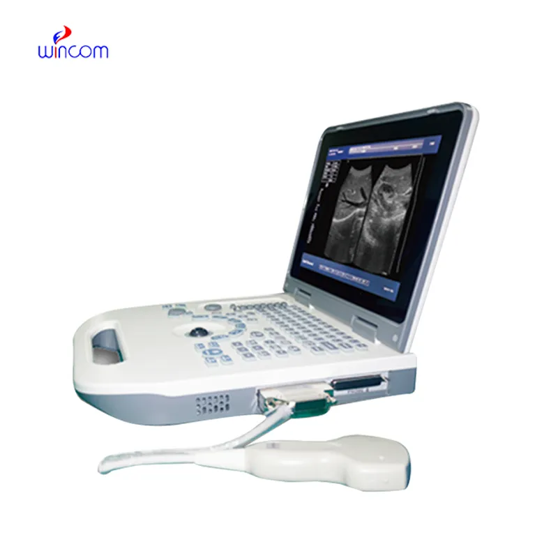
Used in hospitals and clinics, the ge ultrasound machine provides immediate visual feedback for a variety of medical evaluation uses. Converting sound waves into live images, the ge ultrasound machine allows physicians to easily detect abnormalities. The ge ultrasound machine assists with making diagnostic processes safer in addition to improving patient outcomes. It possesses an ergonomic shape alongside digital integration capabilities that support simple data sharing and medical record documentation.
Q: What is the primary function of an ultrasound scannert? A: Ultrasound scanners are designed to create real-time images of internal organs, tissues, and blood flow using high-frequency sound waves. Q: How does the ultrasound scannert ensure clear imaging results? A:It uses advanced converter technology and digital processing to enhance image clarity and contrast. Q: In what medical fields is the ultrasound scannert commonly used? A: It is widely used in obstetrics, cardiology, urology, radiology, and emergency medicine. Q: Is the ultrasound scannert safe for repeated use? A: Yes, it is non-invasive and does not emit radiation, making it safe for frequent diagnostic applications. Q: Can the ultrasound scannert store and share imaging data? A: Yes, it supports data storage, retrieval, and digital transfer for easy integration with hospital systems.
The delivery bed is well-designed and reliable. Our staff finds it simple to operate, and patients feel comfortable using it.
We’ve been using this mri machine for several months, and the image clarity is excellent. It’s reliable and easy for our team to operate.
To protect the privacy of our buyers, only public service email domains like Gmail, Yahoo, and MSN will be displayed. Additionally, only a limited portion of the inquiry content will be shown.
Hello, I’m interested in your centrifuge models for laboratory use. Could you please send me more ...
I’d like to inquire about your x-ray machine models. Could you provide the technical datasheet, wa...
E-mail: [email protected]
Tel: +86-731-84176622
+86-731-84136655
Address: Rm.1507,Xinsancheng Plaza. No.58, Renmin Road(E),Changsha,Hunan,China

 af
af
 es
es
 ar
ar
 tr
tr
 sw
sw
 pt
pt
 th
th
 ur
ur
 bn
bn
 ne
ne
 vi
vi
 km
km
 lo
lo
 de
de
 ru
ru
 fi
fi
 nl
nl
 fa
fa
 fr
fr
 ko
ko