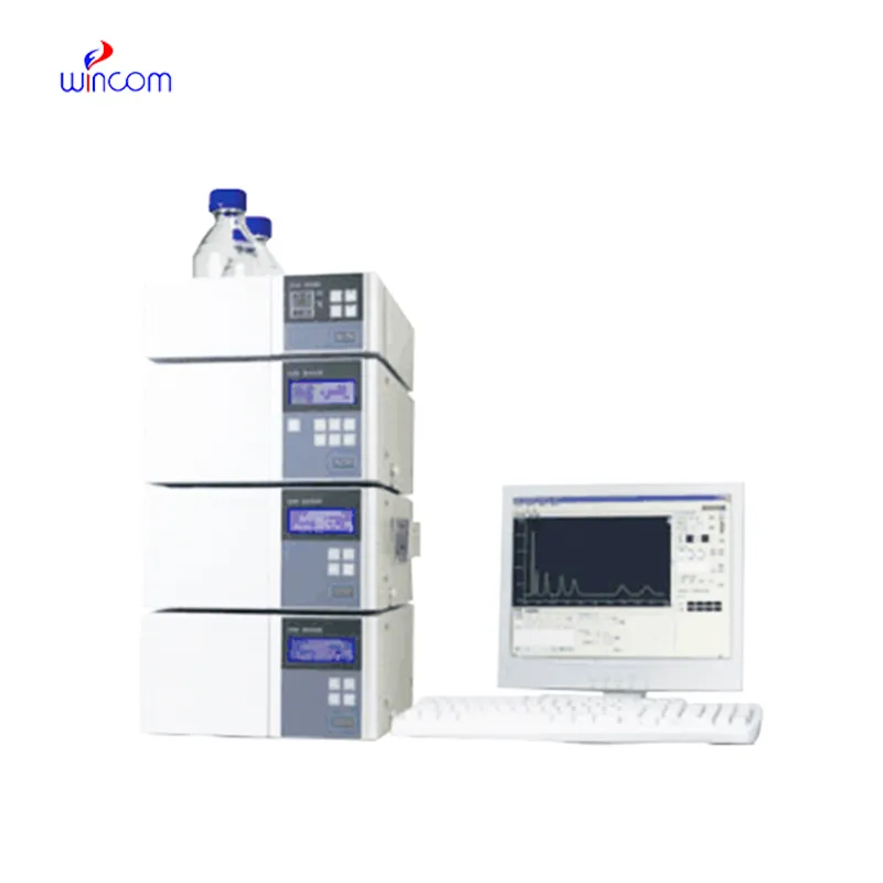
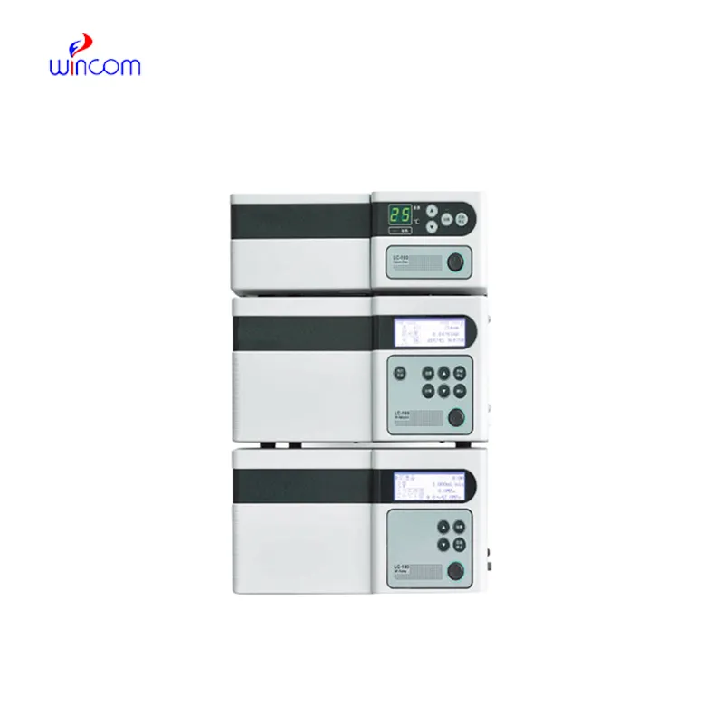
Hospitals and biomed research centers employ high liquid chromatography that help optimize patient testing and lab work. By being able to distinguish, measure, and analyze drugs, metabolites, and biomolecules, high liquid chromatography is a necessary tool in patient testing. Lab professionals incorporate high liquid chromatography into lab work on a daily basis. Reproducibility and analytical ability make high liquid chromatography an irreplaceable tool in assisting with patient testing.

high liquid chromatography allows the personnel of hospitals and laboratories to keep an eye on the presence of environmental pollutants in sterile drugs. It purifies and recognizes the remaining solvents, preservatives, and other possible impurities thus, confirming safety and meeting the requirements of regulatory authorities. This technology is vital in the battle against exposing patients to toxic agents.
The forthcoming breed of high liquid chromatography will put a spotlight on intelligent instruments that are connected with cloud-based surveillance. Through this monitoring, hospitals will be able to gain a remote view of laboratory activities and the results of sample analysis. Lab productivity will be greatly increased by the upcoming high liquid chromatography, and together with the new features, patient testing and therapy monitoring even in difficult clinical settings will be more accurate.
Routine upkeep of high liquid chromatography is of utmost importance in clinical laboratories to maintain the accuracy of patient sample analysis. Regular cleaning of pipes, changing of deteriorated seals and calibration of measuring instruments will block adulteration and keep the latter's sensitivity. Lab personnel must record maintenance activities and keep watch over system performance. Constant attention guarantees that high liquid chromatography provides dependable, reproducible results for hospital diagnosis and research work.
In today's laboratories, high liquid chromatography is indispensable for chemical analysis and serves as a primary instrument. Detection of compounds in intricate mixtures is first done through separation and then identification. Consequently, researchers can precisely check the interactions between molecules. high liquid chromatography is regarded to have extremely high reproducibility and it shares its strength with the fields of pharmaceuticals, biochemistry, and environmental science. Its alliance with sensitive detectors leads to the accurate measurement of very small amounts. high liquid chromatography is the trustworthy partner of lab technicians in validation of experiments, profiling of samples, and development of analytical methods. It not only gives consistent and detailed results but also boosts the efficiency of laboratories and at the same time, makes sure that the data obtained from research is reliable and thus, supports the advanced scientific inquiries that are conducted in various disciplines.
Q: What types of HPLC columns are available? A: Reversed-phase, normal-phase, ion-exchange, and size-exclusion columns are the main types of columns used according to the nature of the analytes. Q: Can multiple samples be analyzed simultaneously? A: Yes, in high-throughput systems, automated sample injection and sequential analysis are among the techniques to achieve this. Q: How does temperature affect HPLC performance? A: Temperature changes can cause variations in separation efficiency and retention times; however, the majority of labs make use of precise temperature control. Q: Can HPLC be integrated with data software? A: Sure, it can be linked with laboratory software for data collection, processing, and reporting. Q: What types of laboratories use HPLC? A: HPLC is employed by hospitals, pharmaceuticals, biochemistry research, and environmental testing labs.
This x-ray machine is reliable and easy to operate. Our technicians appreciate how quickly it processes scans, saving valuable time during busy patient hours.
The water bath performs consistently and maintains a stable temperature even during long experiments. It’s reliable and easy to operate.
To protect the privacy of our buyers, only public service email domains like Gmail, Yahoo, and MSN will be displayed. Additionally, only a limited portion of the inquiry content will be shown.
Could you please provide more information about your microscope range? I’d like to know the magnif...
Hello, I’m interested in your centrifuge models for laboratory use. Could you please send me more ...
E-mail: [email protected]
Tel: +86-731-84176622
+86-731-84136655
Address: Rm.1507,Xinsancheng Plaza. No.58, Renmin Road(E),Changsha,Hunan,China

 af
af
 es
es
 ar
ar
 tr
tr
 sw
sw
 pt
pt
 th
th
 ur
ur
 bn
bn
 ne
ne
 vi
vi
 km
km
 lo
lo
 de
de
 ru
ru
 fi
fi
 nl
nl
 fa
fa
 fr
fr
 ko
ko