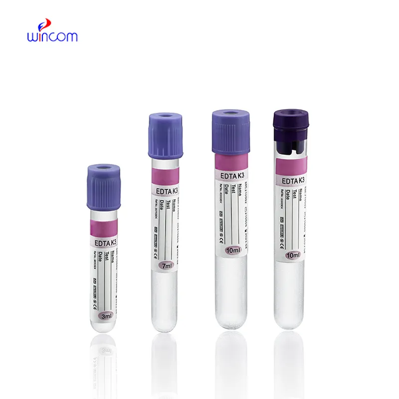
Hospitals gain the advantages of hplc-ms through the precise surveillance of patient's chemical composition and drug concentrations, among other things. The device is capable of separating the different substances with high efficiency, which ensures the delivery of reliable analytical data even if the biological sample is complex. The lab personnel are using hplc-ms for the purpose of continual quality monitoring, which helps in dosage adjustment and patient care. Its accuracy simultaneously supports clinical decision-making as well as research studies, which leads to the laboratories having a versatile and trustworthy analytical platform that can meet the requirements of the healthcare sector and improve the outcomes for the patients.

hplc-ms allows the personnel of hospitals and laboratories to keep an eye on the presence of environmental pollutants in sterile drugs. It purifies and recognizes the remaining solvents, preservatives, and other possible impurities thus, confirming safety and meeting the requirements of regulatory authorities. This technology is vital in the battle against exposing patients to toxic agents.
Advanced software platforms for predictive analytics in healthcare are going to be part of the hplc-ms integration. The hospitals will take advantage of the real-time data provided by the patient samples to influence their clinical decisions. Molecular profiling as well as automated quality control and laboratory efficiency will be thehplc-ms future applications targeting the improvement of patient care.
Preventive maintenance is hplc-ms that play a very important role in clinical and hospital laboratories. The routine performance of flushing columns, cleaning injector valves, and monitoring pressure stability extends the life of the system. The laboratory staff is required to keep records of maintenance activities, replace consumables in a timely manner, and use solvents that are compatible. All of these practices are essential for the instruments' performance retention, lifespan extension, and high-quality analytical results, both in patient sample testing and research.
Therapeutic drug monitoring relies heavily on hplc-ms in hospital settings. It determines the concentration of drugs in the body to guarantee efficiency and security. The laboratory staff uses it for the examination of blood, serum, or urine samples, and signifies small molecular compounds with high accuracy. By yielding consistent outcomes, hplc-ms services the medics in changing the amounts and preventing side effects. Its use goes to hormone level testing, metabolite analysis, and pharmacokinetics research. With quick processing and accurate information, hplc-ms is a part of the hospital patient care, making evidence-based treatment decisions possible and enhancing clinical outcomes in different departments.
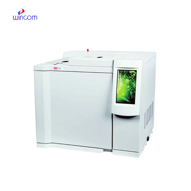
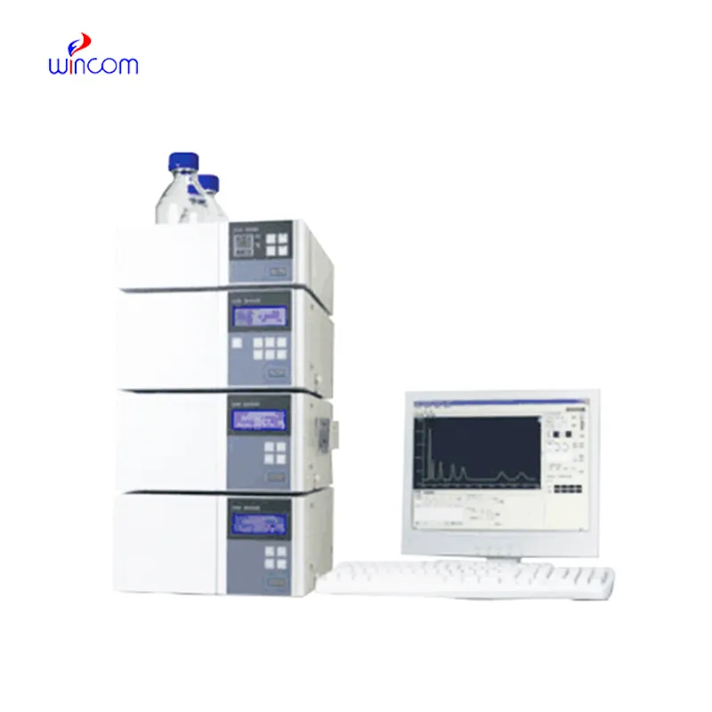
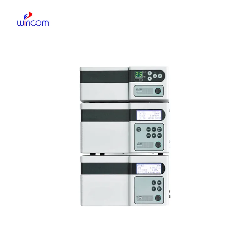
Q: Do you need special training for HPLC operation? A: The answer is yes, training is a prerequisite to accurately and safely using pumps, columns, and detectors. Q: What type of maintenance does HPLC have? A: It requires cleaning, flushing, and inspection of all components as well as calibrating. Q: Is it possible to use HPLC in drug monitoring? A: Sure, it is a common practice in hospitals to monitor the levels of therapeutic drugs and also to identify metabolites in the samples taken from the patients. Q: What is the duration of analysis using HPLC in a typical case? A: The analysis time can range from a few minutes to more than an hour depending on the nature of the sample and the kind of column used. Q: Is HPLC a good choice for environmental testing? A: Yes, it can be used to find out the presence of pollutants, pesticides, and other harmful substances in water, soil, and air samples.
The delivery bed is well-designed and reliable. Our staff finds it simple to operate, and patients feel comfortable using it.
We’ve used this centrifuge for several months now, and it has performed consistently well. The speed control and balance are excellent.
To protect the privacy of our buyers, only public service email domains like Gmail, Yahoo, and MSN will be displayed. Additionally, only a limited portion of the inquiry content will be shown.
We’re currently sourcing an ultrasound scanner for hospital use. Please send product specification...
Could you share the specifications and price for your hospital bed models? We’re looking for adjus...
E-mail: [email protected]
Tel: +86-731-84176622
+86-731-84136655
Address: Rm.1507,Xinsancheng Plaza. No.58, Renmin Road(E),Changsha,Hunan,China

 af
af
 es
es
 ar
ar
 tr
tr
 sw
sw
 pt
pt
 th
th
 ur
ur
 bn
bn
 ne
ne
 vi
vi
 km
km
 lo
lo
 de
de
 ru
ru
 fi
fi
 nl
nl
 fa
fa
 fr
fr
 ko
ko