Built from high-quality optics, the microscope depth of field provides higher clarity for scientific and educational use. The durable body provides stable operation, and the adjustable head and stage setup provide ergonomic convenience. Advanced illumination systems enable observation with high contrast of transparent and reflected samples. The microscope depth of field is compatible with digital cameras and display devices, enabling real-time observation and recording of microscopic structures for further study and analysis.

The microscope depth of field has a wide range of professional and academic uses. In biomedical labs, it is used to analyze cell morphology and identify abnormalities. Industrial scientists rely on the microscope depth of field in testing product consistency, micro defect detection, and surface characterization. In agriculture, it is used to study plant diseases, seed morphology, and pest interactions. Museums and conservation centers apply the microscope depth of field in analyzing artwork materials to ensure proper preservation and restoration of historical works.

The microscope depth of field of the future will be to expand its analytical power. Future models will integrate optical accuracy with the enhancement of the computer, creating hybrid devices with real-time analysis functions. Automation will ease routine operations, making laboratory workflow more efficient. The microscope depth of field will also be able to integrate cloud-based platforms for real-time sharing of data and remote access. Environment-friendly technology development will yield models that are energy-efficient without sacrificing precision but reduce environmental impact.
A well-maintained microscope depth of field gives reliable performance and long operating life. Check optical elements regularly for dust, fingerprint, or oil residue. Use only authorized manufacturer cleaning materials to prevent lens coating damage. Store the microscope depth of field upright, supported, and covered when not in use. Check focusing mechanisms for smooth operation and illumination system for uniform brightness. Standard maintenance procedures minimize downtime and preserve imaging quality for education and research.
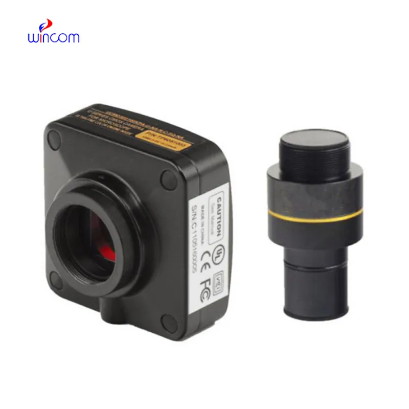
The microscope depth of field bridges the visible and invisible by rendering small particles and organisms visible. Using a lens system and controlled light, the microscope depth of field enables scientists and students to study samples with utmost precision. It has diverse applications in medicine, biology, electronics, and quality control. Digital and fluorescence forms extend study accuracy, simplifying visualization and data recording in most areas of science.
Q: What is a microscope used for? A: A microscope is used to magnify tiny objects or structures, allowing detailed observation of cells, microorganisms, and materials that are invisible to the naked eye. Q: How often should a microscope be calibrated? A: To maintain measurement accuracy and ensure accurate focus during research or analysis, regular calibration should be performed, typically once or twice a year. Q: What type of light source is commonly used in a microscope? A: Most modern microscopes use LED or halogen light sources, which provide stable light and adjustable brightness for clear images at a wide range of magnifications. Q: Can a microscope be connected to a computer? A: Yes, many microscope models feature USB or HDMI ports that allow image capture and digital display through specialized imaging software. Q: How should a microscope be stored when not in use? A: A microscope should be covered with a dust shield and stored in a cool, dry location to prevent contamination and protect optical components from humidity.
The centrifuge operates quietly and efficiently. It’s compact but surprisingly powerful, making it perfect for daily lab use.
This ultrasound scanner has truly improved our workflow. The image resolution and portability make it a great addition to our clinic.
To protect the privacy of our buyers, only public service email domains like Gmail, Yahoo, and MSN will be displayed. Additionally, only a limited portion of the inquiry content will be shown.
Could you share the specifications and price for your hospital bed models? We’re looking for adjus...
Could you please provide more information about your microscope range? I’d like to know the magnif...
E-mail: [email protected]
Tel: +86-731-84176622
+86-731-84136655
Address: Rm.1507,Xinsancheng Plaza. No.58, Renmin Road(E),Changsha,Hunan,China

 af
af
 es
es
 ar
ar
 tr
tr
 sw
sw
 pt
pt
 th
th
 ur
ur
 bn
bn
 ne
ne
 vi
vi
 km
km
 lo
lo
 de
de
 ru
ru
 fi
fi
 nl
nl
 fa
fa
 fr
fr
 ko
ko