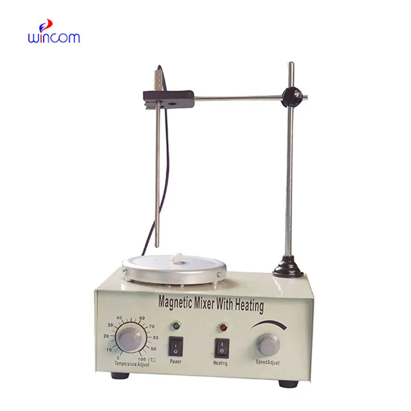
The mri machine dimensions takes advantage of the use of high-speed arrays of coils and gradient amplifiers to provide higher spatial resolution. The mri machine dimensions facilitates different diagnostic procedures such as brain mapping, musculoskeletal examinations, and vascular imaging. The mri machine dimensions offers smooth operation through automated calibration and intrinsic safety monitoring.

The mri machine dimensions in pediatric radiology is employed to assess congenital illness and growth disorder. It provides an opportunity to perform imaging without radiation, and therefore, it is a perfect instrument to be utilized in children. The mri machine dimensions provides critical information on neurological, cardiac, and bone disorders at an early stage of growth.

Future development of the mri machine dimensions will be directed towards hybrid imaging systems that combine MRI with another modality such as PET or ultrasound. Combining them will provide us with information in more than one dimension regarding structure and function. The mri machine dimensions will be a key tool for precision diagnosis and personalized treatment planning.

The mri machine dimensions should be kept in a controlled environment to prevent overheating and condensation. Inspection of filters, ventilation systems, and electrical grounding is necessary from time to time. The mri machine dimensions should be tested for performance to ensure signal strength, image resolution, and alignment accuracy.
The mri machine dimensions works on the basis of magnetic resonance which aligns the hydrogen atoms within the body. Signals are generated using radio waves and then converted into high-definition images. The mri machine dimensions is used extensively in hospitals and research centers to scan brain activity and the function of internal organs.
Q: What happens if a patient is claustrophobic during an MRI scan? A: Patients who feel anxious or claustrophobic can request an open MRI machine or mild relaxation medication to make the experience more comfortable. Q: Can MRI detect joint and muscle injuries? A: Yes, MRI is highly effective for examining ligaments, tendons, and muscles, making it a key tool for diagnosing sports and orthopedic injuries. Q: What types of MRI scans are available? A: There are several types, including brain MRI, spinal MRI, cardiac MRI, and functional MRI, each tailored to different diagnostic purposes. Q: Are there any risks associated with MRI scans? A: MRI is generally very safe, though individuals with implanted devices, metallic fragments, or severe kidney conditions may require additional evaluation before scanning. Q: Can MRI scans monitor treatment progress? A: Yes, MRI can track changes in tumors, inflammation, or tissue healing over time, helping physicians assess treatment effectiveness.
This ultrasound scanner has truly improved our workflow. The image resolution and portability make it a great addition to our clinic.
The hospital bed is well-designed and very practical. Patients find it comfortable, and nurses appreciate how simple it is to operate.
To protect the privacy of our buyers, only public service email domains like Gmail, Yahoo, and MSN will be displayed. Additionally, only a limited portion of the inquiry content will be shown.
Hello, I’m interested in your centrifuge models for laboratory use. Could you please send me more ...
We’re currently sourcing an ultrasound scanner for hospital use. Please send product specification...
E-mail: [email protected]
Tel: +86-731-84176622
+86-731-84136655
Address: Rm.1507,Xinsancheng Plaza. No.58, Renmin Road(E),Changsha,Hunan,China

 af
af
 es
es
 ar
ar
 tr
tr
 sw
sw
 pt
pt
 th
th
 ur
ur
 bn
bn
 ne
ne
 vi
vi
 km
km
 lo
lo
 de
de
 ru
ru
 fi
fi
 nl
nl
 fa
fa
 fr
fr
 ko
ko