The v scanner ultrasound combines state-of-the-art digital imaging algorithms that boost contrast and depth perception, which leads to soft tissues being visualized more clearly. The intelligent interface features customizable scanning modes and user profiles for easier operation. Due to its low power consumption and sturdy construction, the v scanner ultrasound maintains performance that is even in high-demand clinical environments.

The v scanner ultrasound is recognized for its great contribution to the field of surgery and thus is employed frequently in operating theaters for providing intraoperative guidance and ascertaining anatomical targets. It can easily locate areas where fluid has collected, determine the condition of the tissue, and provide evidence that the procedure has been successful. The v scanner ultrasound can also be used dynamically and thus in sports medicine for imaging of muscles and tendons during movement analysis.

The v scanner ultrasound can look forward to getting advantages from miniaturization and wearable technologies. Portable or handheld versions of the v scanner ultrasound will become more widespread to facilitate quick diagnoses in rural as well as emergency setups. The integration of telemedicine services will thus facilitate concurrent consultations via the v scanner ultrasound.

The v scanner ultrasound needs to be maintained based on the manufacturer's recommendations. The transducers should be kept in specialized holders. The cleaning agent should be non-corrosive. The electrical contacts should remain dry. Functional tests should be carried out on a regular basis to ensure that the v scanner ultrasound functions properly and remains a safe instrument.
With the advanced imaging technology, the v scanner ultrasound provides physicians unobstructed and precise images of internal organs. It is employed in the early disease diagnosis as well as in patient tracking. The v scanner ultrasound functions by utilizing sound wave reflections to generate dynamic images, qualifying it as an essential tool in modern medical diagnostics. Through the v scanner ultrasound, fast, non-invasive testing is facilitated for real-time assessment to support clinical decisions.
Q: What makes the ultrasound scannert effective for diagnostic imaging? A: Its high-frequency sound wave technology allows accurate visualization of internal body structures in real time. Q: How portable is the ultrasound scannert? A: The device features a compact and lightweight design, allowing easy movement between clinical departments. Q: What types of probes are compatible with the ultrasound scannert? A: It supports multiple probe types, including linear, convex, and phased array probes for varied diagnostic needs. Q: Does the ultrasound scannert require special training to operate? A: Basic technical training is recommended to maximize its imaging performance and functionality. Q: How long can the ultrasound scannert operate continuously? A: It is designed for extended use with efficient cooling systems and stable power performance.
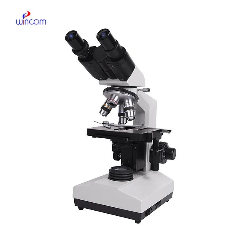
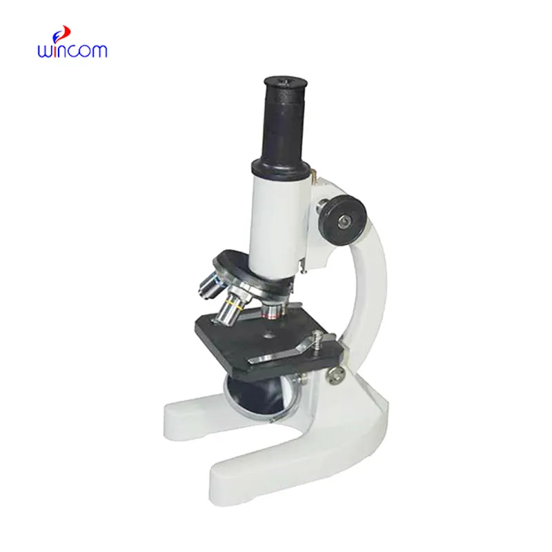
The microscope delivers incredibly sharp images and precise focusing. It’s perfect for both professional lab work and educational use.
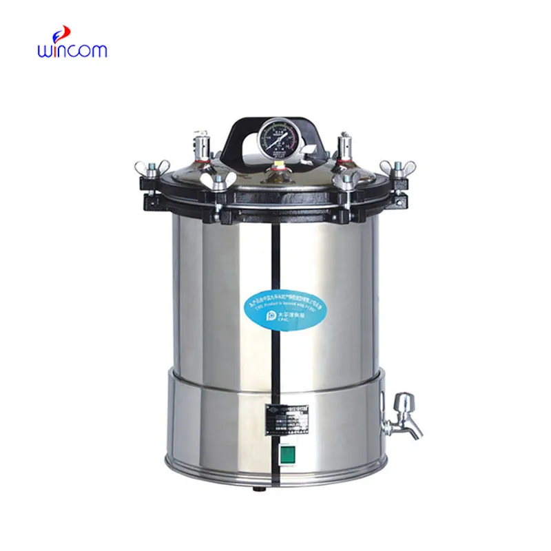
The centrifuge operates quietly and efficiently. It’s compact but surprisingly powerful, making it perfect for daily lab use.
To protect the privacy of our buyers, only public service email domains like Gmail, Yahoo, and MSN will be displayed. Additionally, only a limited portion of the inquiry content will be shown.
I’m looking to purchase several microscopes for a research lab. Please let me know the price list ...
We are planning to upgrade our imaging department and would like more information on your mri machin...
E-mail: [email protected]
Tel: +86-731-84176622
+86-731-84136655
Address: Rm.1507,Xinsancheng Plaza. No.58, Renmin Road(E),Changsha,Hunan,China

 af
af
 es
es
 ar
ar
 tr
tr
 sw
sw
 pt
pt
 th
th
 ur
ur
 bn
bn
 ne
ne
 vi
vi
 km
km
 lo
lo
 de
de
 ru
ru
 fi
fi
 nl
nl
 fa
fa
 fr
fr
 ko
ko